Our Research
The Cardiovascular Calcification Research Laboratory studies the causes of disorders of mineralization in the vasculature, such as calcium mineral deposition in arteries and on heart valves. Calcific disease was previously considered a passive, inevitable process, but it is now known to recapitulate embryonic bone formation under the control of bone differentiation factors and inflammatory mediators, in large part due to our research. Our discoveries include developing the first cell culture model of vascular calcification, isolating the artery wall cell capable of mineralization, and identifying the cell as a multipotential mesenchymal stem cell with substantial self-renewal capacity. We also demonstrated spontaneous formation of periodic patterns by these cells and identified the molecular morphogens responsible and discovered their left-right chirality in migration across a micromachined matrix interface. We proposed the lipid hypothesis of osteoporosis and showed the hyperlipidemia reduces bone density and the response to bone anabolic treatment of osteoporosis. This research is essential for developing the first medical treatment for calcific diseases.
Research Lab Photos
Vascular Tissue Mineralization
Atherosclerotic Calcification
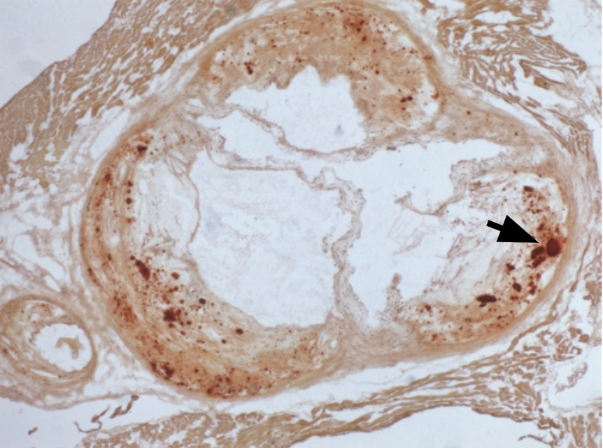
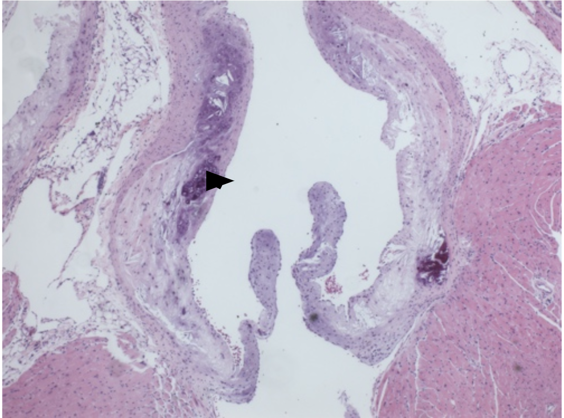
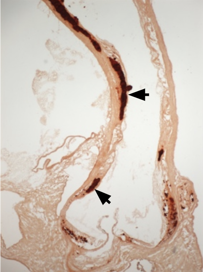
We and others have shown the presence of osteochondrogenic cells (arrowhead) in mice on a high-fat diet develop aortic root calcification (arrows).
Computational Modeling of Calcified Deposits
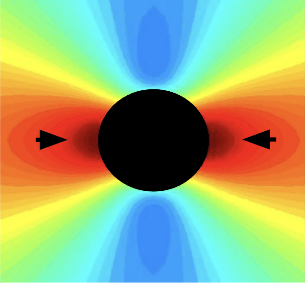
We have demonstrated that calcium deposits are expected to increase rupture stresses (arrows) in the artery wall, based on mechanical and finite element models.
Spontaneously Calcifying Vascular Cells
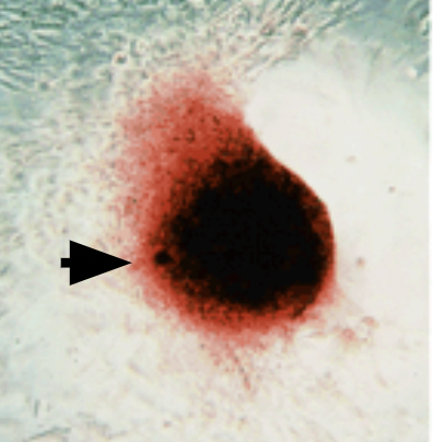
We have shown that, in culture, vascular cells isolated from the aortic media aggregate into nodules (spheroids, arrow) and produce hydroxyapatite mineral.
Spontaneous Calcific Nodule Formation
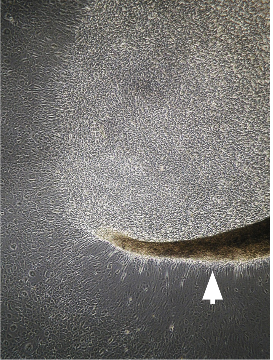
We have discovered that calcifying vascular cell aggregation into nodules/spheroids (arrow) is enhanced by transforming growth factor-beta (TGF-b) as well as by tumor necrosis factor-alpha (TNF-a).
Lipids & Calcification
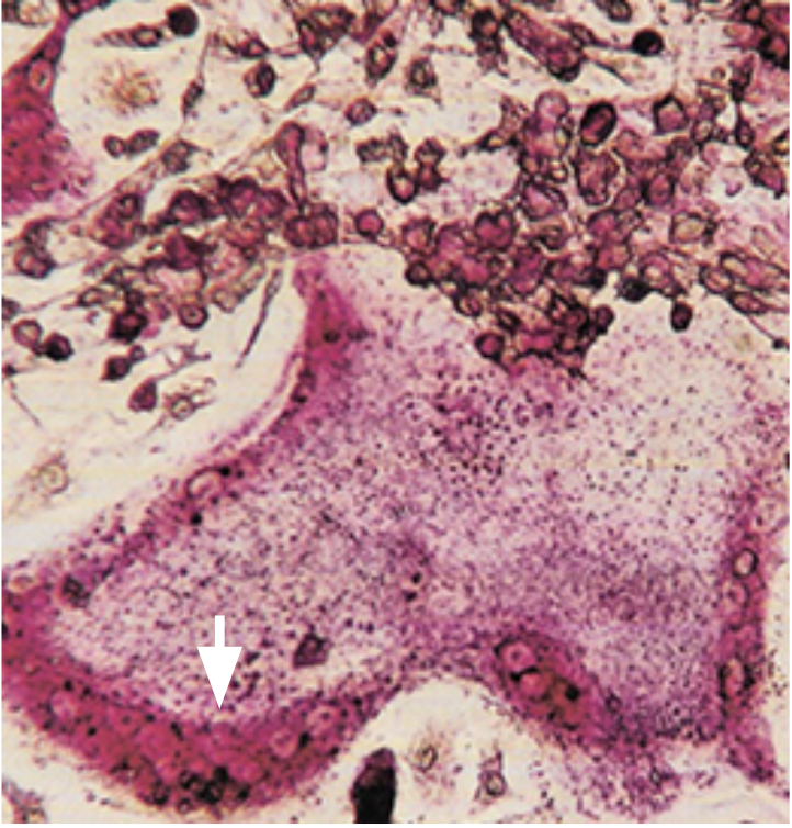
We have introduced the Lipid Hypothesis of osteoporosis, showing that bioactive phospholipids that trigger cardiovascular calcification also promote osteoporosis, inhibiting differentiation of bone forming osteobalsts while promoting differentiation of bone resorbing osteoclasts. Light microscopy of bone-marrow derived a multinucleated (arrow) osteoclast stained for tartrate-resistant acid phosphatase (TRAP).
In Vivo Molecular Imaging of Cardiovascular Calcification
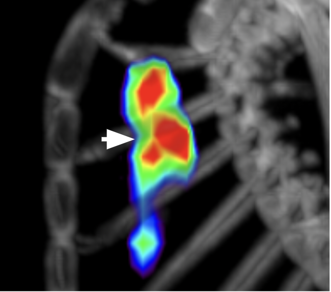
We have demonstrated that 18F-NaF positron emission tomographic (PET) labels cardiovascular calcification (arrow) in mice. Fluoride ions adsorb onto surfaces of hydroxyapatite, replacing the hydroxyl groups, to form fluoroapatite. Microarchitectural changes of cardiovascular calcification are affected in response to interventions, such as exercise, aging, and lipid-lowering drugs.

